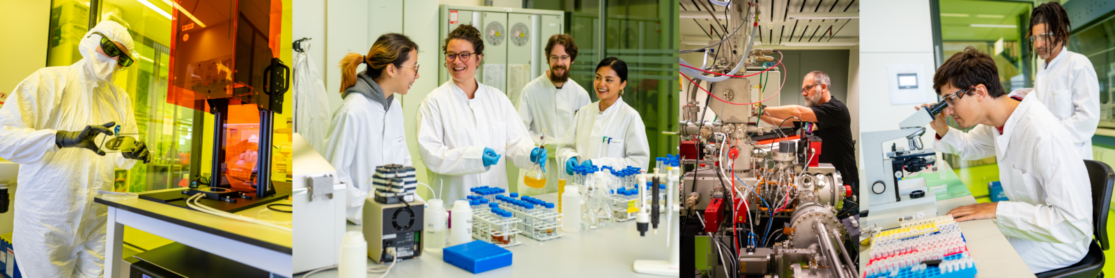
Advantages & limitations
 NanoSIMS is a versatile analytical technique. It has key advantages but also comes with some practical limitations, which should be kept in mind when considering to use NanoSIMS.
Key advantages include:
-
Lateral spatial resolution of ~50 nm is achieved using a highly focused primary ion beam.
-
Imaging capability is achieved by rastering the primary ion beam over the sample surface. For this type of measurements, supplementary information is crucial to allow navigation on the sample. This information can include images obtained via Scanning Electron Microscopy (SEM), Transmission Electron Microscopy (TEM) or Fluorescence Microscopy with markings or coordinates.
-
The high mass resolution of M/dM>5000 allows discrimination between ions of very close masses (e.g., 13C and 12C1H).
-
Samples prepared for Transmission Electron Microscopy (TEM), Scanning Electron Microscopy (SEM), Electron Microprobe (EPMA) and Fluorescence Microscopy are usually suitable for the analysis by NanoSIMS.
-
In principle, atomic or molecular ions with masses ranging from hydrogen to uranium and beyond can be detected by NanoSIMS.
-
Noble gases cannot be detected as they do not ionize under the conditions used by the instrument.
-
The NanoSIMS 50L model offers simultaneous detection of up to 7 masses. However, there are some restrictions due to the geometry of the instrument and differences in ionizability among elements:
-
During simultaneous analysis, the minimum and maximum masses cannot differ by more than a factor of 22. Some elements are very difficult to ionize and are therefore preferably detected as molecular ions (e.g., N is detected as CN–). This brings along problems with interferences.
-
There is a technical limit regarding the separation of masses that can be detected simultaneously. An approximate formula for calculating the difference in masses that can be detected simultaneously is ΔM=0.017 ∙ √ (Mmax ∙ M), where Mmax is the maximum detected mass and M is the base mass for which one wants to simultaneously detect also the mass M + ΔM.
-
During ionization, some elements are preferably ionized as positive ions and others as negative ions. Therefore, it is possible to simultaneously detect only a combination of elements that ionize either as positive or negative ions.
-
Related to the previous item, negative secondary ions are detected using the positively charged Cs+ primary ion beam, while positive secondary ions are detected using the negatively charged O– primary ion beam. Although both of these primary ion sources are available, only one can be used at a given time. As NanoSIMS is a destructive technique, this limitation implies that negatively and positively charged secondary ions cannot be measured from the very same sample volume. Although they can be measured in sequence, switching between primary ion sources is a rather lengthy procedure imposing constrains on the experimental design and the subsequent NanoSIMS analysis.
-
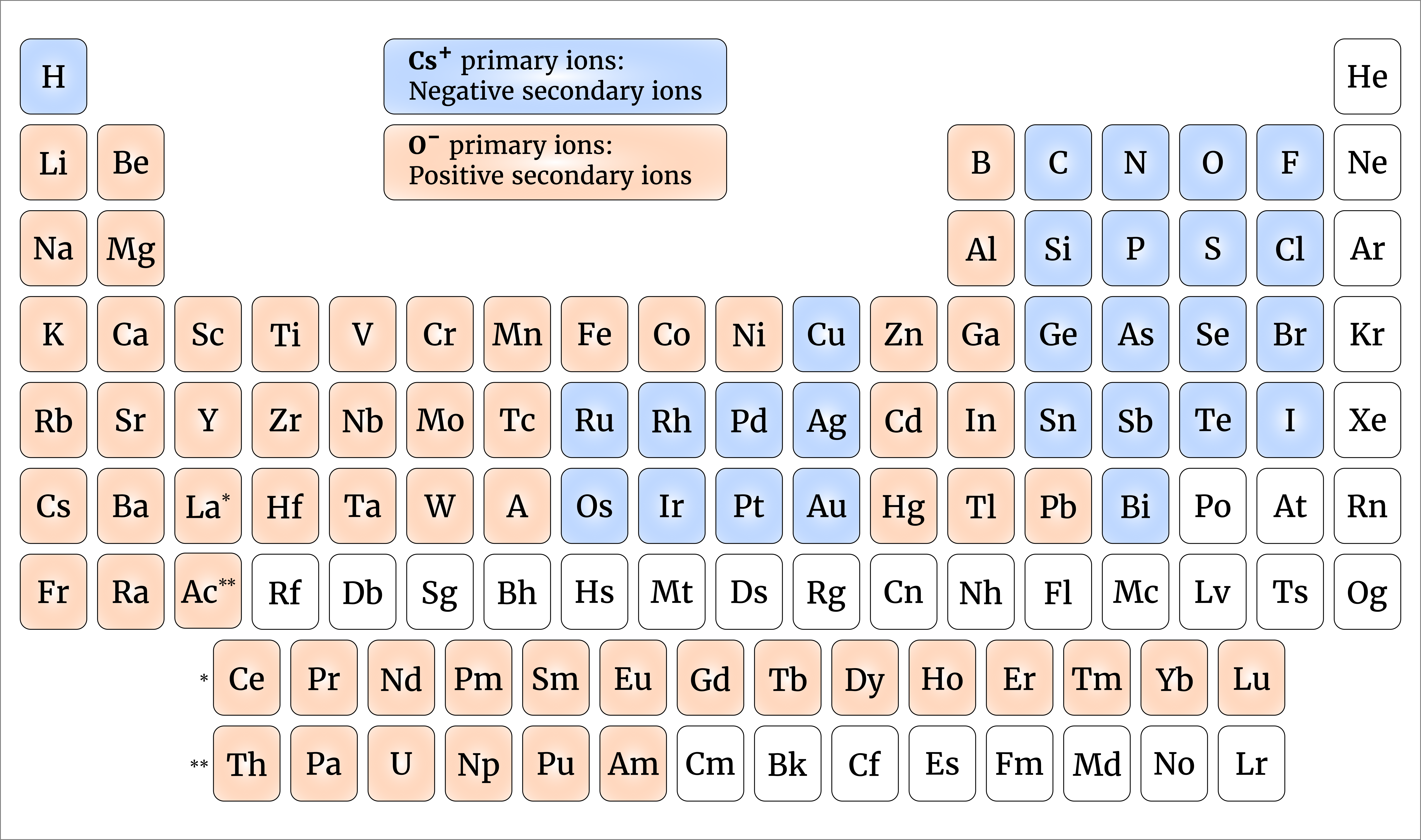
Depending on the position in the table, elements can be detected by NanoSIMS as either positive or negative ions.
Practical limitations include:
- NanoSIMS is a destructive analytical technique, as the sample surface is eroded during the measurement. If combined with other imaging techniques, NanoSIMS should preferably be used as the last analytical step.
-
NanoSIMS is typically a rather low through-put measurement, taking tens of minutes up to several hours to acquire an image with a sufficient resolution and precision.
-
To achieve maximum sensitivity and precision of the NanoSIMS instrument, components of the ion optics need to be accurately aligned. Besides frequent fine-tuning, this requires adjustments when moving from one field of view on the sample to another or when changing between samples. Therefore, the total analysis of a batch of samples may take several days of continuous measurements.